HeadWorks Physiotherapy
  • Home
  • Temporomandibular Joint
    • Temporomandibular Disorders (TMD)
    • Causes of TMD
    • Symptoms of TMD
  • Orofacial Myofunctional Therapy
  • Physiotherapy
    • What to Expect
    • Your Visits and Fees
  • Blog
  • About
  • Contact

Some notes on pain

23/2/2016

0 Comments

 
I am writing down some (random) notes on pain that I have gleaned from various lectures and readings...organising my thoughts:
  • Any credible evidence of danger to your body will modulate pain.
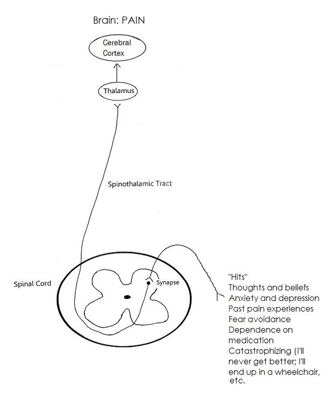
  • Related tags share neurons (neurotags). Take back pain for example:
  1. Beliefs about your back
  2. Thoughts and feelings about your back
These thoughts and beliefs share the same brain cells, and when activated (e.g., when you think about your back), may increase the excitability of your back pain, may lead to catastrophic interpretation ("My back pain will never get better").

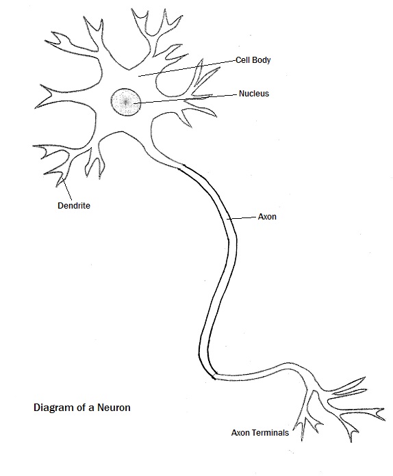
  • Neurons that fire together, wire together.
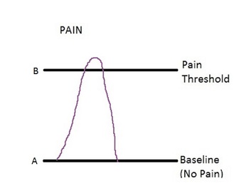
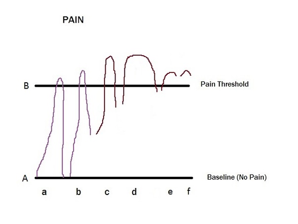
  • When pain persists:
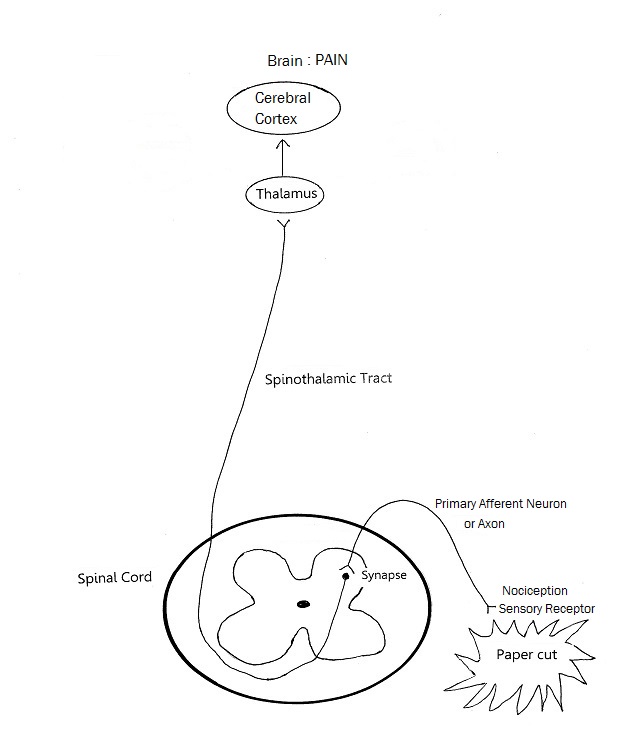
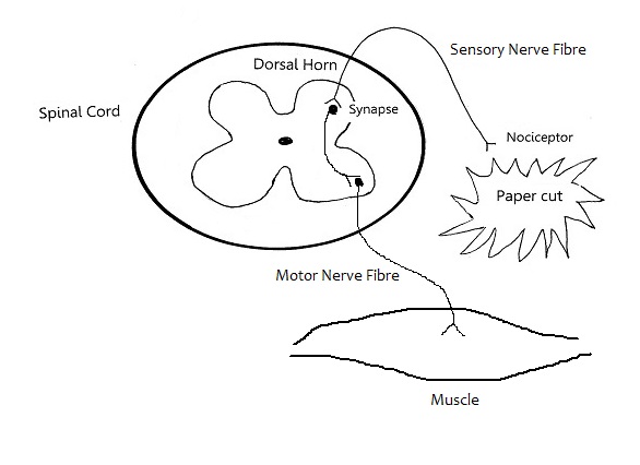
  1. there is increased sensitivity, i.e., the neurotag that produces your back pain is more sensitive, such that even just watching someone pick up a box would make your back hurt (this pain has been picked up in brain scans, so we know that subjects are not lying about their pain) --the network or pain pathway, if you like, becomes stronger and stronger. (The more you feel pain, the better your body becomes at producing it--it is reinforcing the neural pathway to producing pain). It is the familiar road with the well-trodden path, it is "all roads lead to Rome" (pain). Sometimes thoughts, sometimes smells, foods, images, sounds, etc can lead to pain. They share the same neurotag that results in pain.
  2. there is decreased precision: the area over which you feel pain becomes diffuse, and hard to pinpoint.


0 Comments

Checklist for orofacial myofunctional disorder

22/1/2016

1 Comment

 
If you answer "yes" to 3 or more questions, you may need to seek the service of an Orofacial Myofunctional Therapist:

  • Do you have an open mouth at rest or mouth breathe?
  • Does your tongue rest or brace against your teeth?
  • Have your teeth moved after orthodontic treatment?
  • Do you often have headaches?
  • Does your jaw or neck often hurt?
  • Are you a stomach or side sleeper?
  • Do you have oral habits such as nail biting, pen chewing, lip licking or chewing, or digit sucking?
  • Do you have a forward head posture?
  • Do you have a lisp at times with saying the "s" sounds?
  • Do you feel that your tongue comes forward when you swallow?
  • Do you drool?
  • Do you have bloating or stomach distress after eating?

Please read this to find out more about Orofacial Myofunctional Therapy (OMT). You can also go here for some background history on OMT.
1 Comment

Why does it still hurt?

7/12/2015

0 Comments

 
Central sensitisation
This is when the nervous system becomes over sensitive to stimulation. The threshold for Pain/sensation is lowered, and it takes less and less provocation to elicit a Pain response.
Because the central nervous system (CNS) is in such a state of high reactivity, a stimulus such as touch or temperature may be interpreted by the brain as pain. This is known as allodynia.
When a very minor painful stimulus, such as a slight bump on the elbow, is perceived as excruciating pain, this is known as hyperalgesia.


Picture
Different stimuli such as light, sound, odours or even thoughts, may trigger allodynia or hyperalgesia.
 
Why does this happen?
Central sensitisation is what develops and maintains chronic pain. It plays a key role in many different chronic pain disorders, such as neck and back pain, whiplash injuries and injuries sustained in a motor vehicle accident, tension headaches, migraine headaches,  fibromyalgia, chronic fatigue syndrome, and irritable bowel syndrome.
Broadly speaking, there are two types of factors that cause central sensitisation:
  1. Predisposing factors: these are factors that exist before pain has occurred, and can be biological, psychological or environmental.
Biological or genetic factors, such as low pain thresholds may predispose to pain chronification. More research is needed to find a causative effect of low pain thresholds on the development of central sensitisation.
Psychological factors like stress have been shown to lower pain thresholds. Stress and anxiety about pain leads to higher pain sensitivities (lower pain thresholds). If the CNS is already over reactive, central sensitisation may be more likely to happen when pain occurs.
  1. Factors after the onset of pain which may lead to central sensitisation:
Conditions such as depression, fear-avoidance (fear of pain and avoidance of activity) and anxiety may develop with the onset of pain. These responses would increase the reactivity of the CNS and lead to central sensitisation.
Chronic pain sufferers may experience non-restorative sleep, which, in turn may cause increased sensitivity to pain: a vicious cycle.
0 Comments

Pain Threshold and "Hits"

4/11/2015

0 Comments

 
Picture
Suppose line A is your baseline where there is no pain, everything is hunky dory, and line B is your pain threshold, beyond which you feel pain.


Picture
In many cases, or at least initially anyway, when we get a “hit”, we get to the point beyond our pain threshold where we feel pain. After a while, when things heal and get better, most of the time we get back down to our baseline of “no pain”. Usually this does not take too long. This is (a) in the picture below.
“Hits” may be physical, as in the form of an injury, accident, disease. They may also be emotional or psychological, as in trouble in relationships, or stress, anxiety, anger, depression, although, the last few may also feed into the whole pain loop in a vicious cycle.    
Over time, as we get more hits, we may not come all the way back to our “no pain” baseline (b), such that when we get hit the next time, it takes less of a hit to get us beyond our threshold (c). Then we might get a really big hit that takes us a long time to recover from (d), only we don’t really fully recover. We might be painfree, but we may be emotionally more fragile, for example.
The next time we get a hit, it may put us over the threshold and then keep us there (e), such that it would take just a tiny hit and we feel pain (f), or, we are in constant (chronic) pain.

Having said that, however, not everybody stays in the chronic pain zone. We will talk more about what keeps some people there in the next post.

It’s like our body accumulates pain (hits).


0 Comments

Orofacial Myofunctional Therapy

20/9/2015

1 Comment

 
This is for people who like to read...there is a lot of information....

So I am going to do a sideways jog and change the subject for this post. I was going to post a blog on Central Sensitization, but this topic has been occupying the better part of my waking hours since I took the course at the end of August. I wanted spend some time organising my thoughts around this topic, and decided to create a new page on the HeadWorks website. Please feel free to head over to this page and have a read. It's interesting stuff. Sorry it's real wordy. I'll add some pictures later.

Orofacial Myofunctional Therapy is an exciting, emerging field of therapy that is involved in the neuromuscular retraining of the oral and facial muscles. It was actually first introduced in the 1900s by an orthodontist (Dr. Alfred Rogers) who experimented with facial muscle exercises.

Brief overview of history:
  • In 1918, Dr. Rogers published: "Living Orthodontic Appliances", in which he cited that muscle function alone would correct malocclusion with no need for retention. (Can you imagine the controversy?)
  • In the early 1900s, Dr. Benno E. Lischer first coined the term "Myofunctional Therapy" after studying with Dr. Rogers.
  • In 1907, Dr. Edward Angle, an orthodontist, wrote articles on the effects of habits. He is responsible for classifying occlusion (Class I, II, III malocclusion).
  • In 1925, Dr. Harvey Stallard studied 7000 children on sleep position and malposed tooth buds and concluded that, "Sleeping on the face during a child's formative years could create malocclusion."
  • 1924-1940: Truesdell and Truesdell were the first to understand the relationship between swallowing anomalies and dysmorphosis. They advised patients to swallow in occlusion. They also felt that atypical swallowing was related to hypertrophy of the tonsils.
  • An orthodontist, Dr. Klein is often quoted: "Living bone is extremely susceptible to the guidance and influence of pressure and stimuli.” (Klein, 1951)
  • In the 1950s, speech therapists began to look at nasal obstruction, oral habits, swallowing, and how they affected speech.
  • In 1960, Dr. Walter Straub had a theory that bottle feeding caused the "perverted" swallow. He developed a series of exercises to correct the swallow, and lectured with 500 cases on record. Dr. Straub wrote many articles published in the American Journal of Orthodontics.
  • There are several other pioneers who developed exercises for successful therapy.
The past helps us understand the present and look to the future.


1 Comment

Descending Inhibition

2/9/2015

4 Comments

 
Picture
So the cannibal tribe is chasing you, and you trip, fall and sprain your ankle. We know that sprained ankles are extremely painful, but if you stop to deal with your sprained ankle, the cannibals will catch you and eat you.

Your brain is going to decide that staying to nurse your sprained ankle will reduce your overall probability of survival, so it shuts the pain off. How does it do that?

1)      Your brain decides that pain is going to reduce your survival rate, so it tells the hypothalamus, “Let’s not have any pain.”

2)      The hypothalamus communicates with the Peri-Aqueductal Gray Matter (PAGM), which is the gray matter surrounding the cerebral aqueduct in the midbrain. The PAGM coordinates the body’s analgesic system. (When the PAGM is electrically stimulated, pain is shown to be eliminated or reduced).

3)      When the PAGM is activated, it sends analgesic impulses down through the brainstem, through the raphe nucleus, which synapses with a descending neuron that goes down one side of the spinal cord (dorsolateral tract) to influence analgesia at the appropriate level of the spinal cord.


As previously described in earlier posts, a nociceptive impulse comes into the dorsal horn of the spinal cord and synapses with a second order neuron to go in the spinothalamic tract to the brain for pain to be recognised/registered.

If it is inappropriate to feel pain, analgesic impulses come down the dorsolateral tract, and at the appropriate level of the spinal cord, a neuron (analgesic neuron) will project from the dorsolateral tract into the area of the synapse between the axon of the sensory neuron and the start of the second order neuron (which would take the nociceptive impulse to the brain).


Picture
This analgesic neuron contains chemical transmitters which fit into the pre- and post- synaptic receptor sites. When these chemical transmitters bind with the pre- and post- synaptic sites, they cause pre- and post-synaptic inhibition respectively, resulting in the whole synaptic area being turned off, inhibiting ongoing propagation of nerve impulses. Therefore, pain is not felt, as the impulse going to the brain has been blocked.

Picture
Interesting to note: these chemical transmitters are opioids. The body produces its own opioids, endorphins, enkephalin substances.  Also interesting to note that the seed of the poppy plant has the same chemical properties as your body’s endogenous opioids. Morphine, when injected, will have the same effect of pain inhibition as these endogenous opioids.

So, when it is not appropriate for you to feel pain in order to ensure your survival , your body activates its own analgesic system, releasing its own opioids, blocking off pain. All this happens outside your awareness.

This is your amazing body.

4 Comments

August 12th, 2015

14/8/2015

0 Comments

 
     Modulation

This is when the brain processes the nociceptive information coming in and assesses how threatening it is to the body.  It needs to answer:

1)      What does this mean

2)      What will be advantageous

Taking Bethany Hamilton (again) for an example, the answer to the first question was obvious.  What would have been advantageous was that she got out of the water as fast as she could (which she did) and got the help she needed as soon as possible. If she had stayed in the water, she would have bled to death, never mind getting the shark in a frenzy. This is called descending inhibition (this will be discussed in a later blog post).

Any credible evidence of danger to your body will modulate Pain. This time, for her, the danger was not the shark bite, but, rather, the danger of bleeding to death in the water. Therefore, no Pain, or minimal Pain.

Picture
The next time she got in the water, she may well have excruciating pain from a scratch by a piece of seaweed. This is because Pain depends on how much your brain thinks you are in, not how much danger you are really in. So, the input from the scratch by the piece of seaweed would have travelled up into the thalamus, routed to the cerebellum, frontal cortex, limbic system. The situation would be assessed by these parts of the brain, and, drawing from the previous experience of the shark bite and nearly bleeding to death, the brain would have sent out a great Pain output, as this time, the meaning would be different.

0 Comments

Terminology continued

20/7/2015

2 Comments

 
In the last blog post, I introduced a few pain terms and basic neuroanatomy. Here are a few more:

1)      Suffering: this refers to a person’s reaction to his pain perception. How and how much a person suffers is influenced by several factors such as his thoughts and beliefs, past pain experiences, how harmful he thinks the injury may be or may become, what kind of attention is drawn to the injury. This is why suffering may not be proportional to the nociception. Remember Bethany Hamilton (May 25, 2015 Blog post).

2)      Pain behaviour is how a person’s suffering is communicated to others. This may be done through audible or visible actions. This is the pain information that is related, narrated or demonstrated by the person.

3)      Transduction: this happens in the peripheral tissues when noxious stimuli are converted into electrical activity in the sensory nerve endings.

4)      Sensory nerve endings: these are the ends of peripheral nerves; dendrites which lie far from the cell bodies of nerves.

5)      Conduction:  this is when the neuron carries the noxious information into the central nervous system.

6)      Transmission: this is when nociceptive information is conveyed from one neuron to another through junctions called synapses. The primary afferent neuron carries the information to the spinal cord, where it synapses with a second-order neuron, which then carries the information on to the higher centres. This input or information is then routed to different areas of the brain (thalamus, cortex, limbic system)

7)      Synapse: this is a junction across which a nerve impulse is passed from an axon terminal to another neuron, muscle cell, or gland cell.

8)      Perception: the recognition and interpretation of pain by the brain. This is the beginning of suffering and pain behaviour, and it varies greatly between people. 

Picture
2 Comments

Terminology

8/7/2015

2 Comments

 
Picture
I thought I’d do something lighter and less convoluted over the summer before going on to “Why it Still Hurts”

Here are a few pain terms and basic neuroanatomy that you might encounter:

1)      Nociception: this is the noxious stimulus which originates from the sensory receptor. Nociception is carried to the Central Nervous System by the primary afferent neuron.

2)      Sensory receptors: these are at the ends of sensory nerves. They receive physical or chemical stimuli.

3)      Central Nervous System (CNS): this is made of the brain and spinal cord. Sensory impulses are transmitted to it and motor impulses pass out from it. It coordinates the activity of the entire nervous system. It is able to modulate noxious stimulation. This is important as it is this modulation which increases or decreases the perception of pain.

4)      Primary afferent neuron: this is a nerve that transmits impulses from receptors to the central nervous system. Efferent neurons transmit signals from the CNS.


​5)     
Axon: this is a nerve fibre. A bundle of many axons form a nerve. This can be sensory or motor.

6)      A neuron is a nerve cell.

7)      Pain: this is the sensation perceived in the cortex resulting from incoming nociceptive input.

8)      Cortex: This is the cerebral cortex, which is the brain’s outer layer of nerve tissue in humans. The cerebral cortex is gray matter. This is where most sensory information is routed to (via the thalamus).


Picture
2 Comments

Back to basics, continued

22/6/2015

0 Comments

 
Picture
The Withdrawal Reflex

In the last blog, I described what happens when you get a paper cut, how the pain signal is transmitted to your brain for you to feel the pain.

Here is a modified diagram from the last post, showing what happens even before the signal reaches your brain:


Before you were even aware of the pain from the paper cut, you probably reacted involuntarily, e.g., pulled the piece of paper away from your finger, or pulled your finger away from the paper, or both. This is a reflex response, called a withdrawal reflex, and happens at the spinal cord level (a spinal reflex) cord to sudden pain. Motor nerve fibres (motor neurons) are activated to bring about arm muscle contraction, to pull away.


This reflex helps to protect the body from damaging stimuli.

The same thing happens when you touch a hot stove. You immediately whip your hand away without even thinking, and before the pain sets in.
Without this spinal reflex, you might leave your hand there until you feel the pain before you removed your hand, at which point you may have severely burnt your hand.

This is your amazing body.


0 Comments
<<Previous
Forward>>

    Author

    Sharon is a physiotherapist focusing her treatment on TMDs and related orofacial and craniofacial pain.

    Archives

    January 2021
    January 2018
    December 2017
    November 2017
    September 2016
    August 2016
    July 2016
    June 2016
    May 2016
    April 2016
    February 2016
    January 2016
    December 2015
    November 2015
    September 2015
    August 2015
    July 2015
    June 2015
    May 2015

    Categories

    All

    RSS Feed

                                                                            www.headworksphysio.com
                                                                                              copyright 2014
Photo used under Creative Commons from jelene